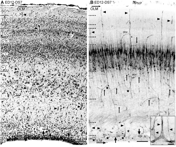
Figure 11. Radial organization of the OT vasculature during DS7 (ED12, HH38) (A: H-E; B: Diaphorase).
(A) There is a remarkable thickening of the SO and the retinorecipient layers of the SGFS. (B) Significant changes in the vascular pattern accompany the retinorecipient layers remodeling and the late differentiation of the C “SGC”. Several different patterns of distribution of collateral branches and tangential to oblique anastomoses can be seen associated to the different TCCs. Inset: a new population of slender straight and radially ascending branches arises from the tangential vessels of the periventricular plexus (thick arrows). acv: ascending branches; dcv: descending branches; thin arrows: anastomoses; asterisk: bifurcations; arrowhead: radial vessel. The densely diaphorase-stained bands located halfway between the ventricular and the pial surface correspond to the layers of NOS+ neurons that typically populate the “i”—“j” complex at this stage [28] (Bars: A,B: 50 µm; Inset: 10 µm)

