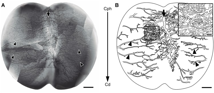
Figure 12. Tangential organization of the perineural plexus (ED7; HH31) (A: Diaphorase).
(A) Image obtained by assembling several partial images of the OT leptomeningeal plexus. The superficial (venous) and the deepest (arterial) plexuses appeared overlapped. (B) Drawing of the largest vessels observed in “A”. The main venous vessels, running along the midline (arrows), and the main leptomeningeal arteries, running in a lateral-to-medial direction (arrowheads) exhibit a higher development in the cephalic region revealing the existence of a cph-cd angiogenic gradient (large arrow in the middle of both figures). The arrows and arrowheads shown in A and B allow the identification of the same vessels in both figures. Inset: Magnification of the box shown in figure “B” illustrating the complexity of the leptomeningeal capillary bed. (Bars: A,B: 500 µm; Inset: 100 µm).

