
Figure 1.
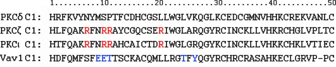
Amino acid sequence alignment of the C1b domain of PKCδ, the C1 domains of PKCζ, PKCι, and hVav1. The residues in PKCζ/ι largely responsible for the lack of phorbol ester binding are shown in red.17 The residues in Vav1 largely responsible for the lack of phorbol ester binding are shown in blue.18