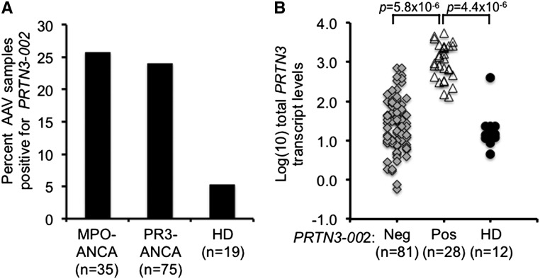
Figure 4.
Frequency of PRTN3–002 mRNA and relationship to total PRTN3 transcripts in peripheral leukocytes. (A) Patients with AAV (n=112) and healthy donors (n=19) were screened for PRTN3–002 mRNA by RT-PCR. Percentage of patients positive for PRTN3–002 mRNA is shown for two serotypes: MPO-ANCA and PR3-ANCA. An ANCA-negative sample and a dual positive ANCA sample were excluded. (B) Total PRTN3 transcript levels determined by TaqMan quantitative RT-PCR, which uses primers in exons 4 and 5, were compared from patients that were negative (Neg; gray diamonds; n=81) or positive (Pos; open triangles; n=28) by RT-PCR for PRTN3–002 mRNA and healthy donors (HDs; black circles; n=12). There were no quantitative RT-PCR data for three patient samples and seven healthy donor samples. P values were calculated by t test.

