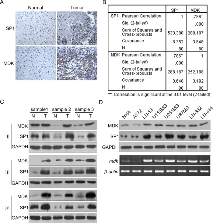
FIGURE 1:
SP1 overexpression correlates with up-regulation of MDK expression in human glioma samples and cell lines. (A) Immunohistochemical staining with specific anti-SP1 and anti-MDK antibodies on glioma tissues. Normal, brain tissues surrounding the tumor; Tumor, human glioma tissues. (B) We quantitatively scored the tissue sections according to the percentage of positive cells and staining intensity as described in Materials and Methods. We then combined the percentage and intensity scores to obtain a total score (range, 0–8). SP1 expression levels correlated positively with MDK expression levels in glioma samples (Pearson's r = 0.79; p < 0.001). (C) SP1 and MDK protein expression levels for three paired low-grade astrocytoma (II), three paired anaplastic astrocytoma (III), and three paired glioblastoma (IV) frozen tissues as determined by Western blot analysis. (D) The SP1 and MDK protein levels in NHA and various glioma cell lines as determined by Western blot.

