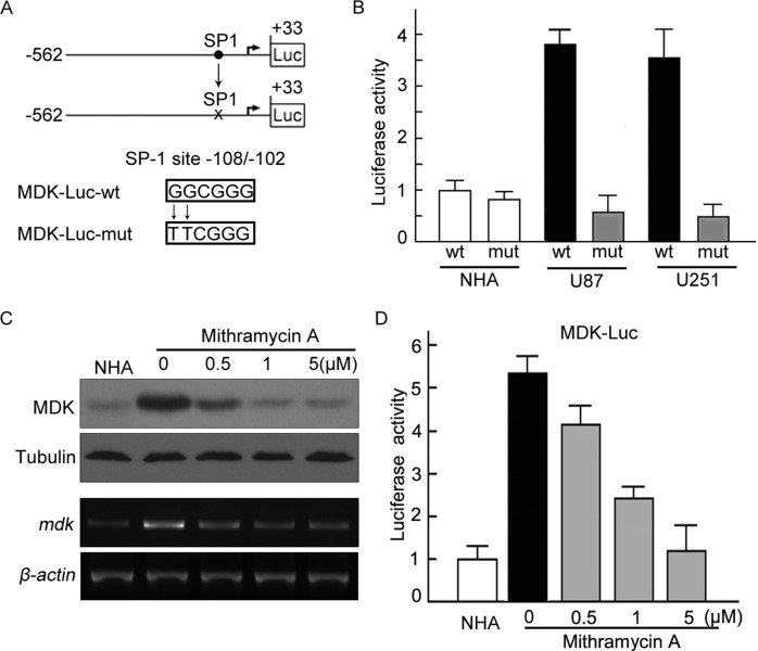
FIGURE 4:
Mutation of the SP1 site abolished activation of the MDK promoter. (A) The core regions of MDK promoter were analyzed by Patch and MatInspector, and the putative conserved SP1 binding site was located at −108/−102. The wild-type SP1 site, GGCGGG (WT), was mutated to TTCGGG (Mut). (B) NHA, U87, and U251 were cotransfected with MDK-Luc wt or mdk-Luc mut, together with pRL-TK for 24 h, respectively. Luciferase activity was measured. (C) Mithramycin A attenuated the up-regulation of MDK in a dose-dependent manner. U87 cells were placed in medium in the absence or presence of mithramycin A at the indicated concentrations for 12 h. MDK expression was assessed by Western blot and RT-PCR (left). (D) Mithramycin A attenuated the activity of mdk promoter. mdk promoter activities were measured by luciferase assay. The experiments were repeated three times, with duplicates for each treatment.

