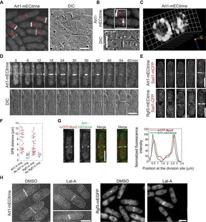
FIGURE 2:
Localization of Art1 to the contractile ring and the septation site during cytokinesis. (A–C) Micrographs of Art1-mECitrine (JW2674) localization at the contractile ring (arrowheads) and septal disk (arrows). (C) Three-dimensional projection of the cells in B imaged with 0.2-μm z-spacing. 1 grid = 0.95 μm. (D) Time course of Art1-mECitrine (JW2674) shows its appearance, constriction, and septal localization. (E and F) Micrographs (E) and quantification (F) of the arrival of Art1 (JW2694) and Rgf3 (JW2748) at the division site using SPB protein Sad1-mCFP as a cell cycle marker. (G) Art1 locates outside of Myo2 in the contractile ring. Micrographs (left) and line scans of fluorescence intensity at the contractile ring (right) in cells expressing both Art1-mECitrine and mCFP-Myo2 (JW4427). The central focal plane was used. (H) Actin-filament independence of Art1 (JW2674) and Rgf3 (JW1105) localization. Scale bars: 5 μm.

