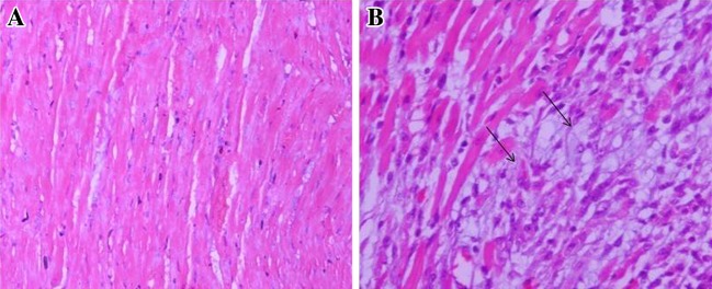
Fig. 8.

Photomicrographs of the heart tissue, a Microscopic section of normal control group rat heart showing normal architecture of cardiac muscle (H&E 100×); b ISP treated group rat heart showing extensive myocardial necrosis and inflammatory cell infiltration (H&E 100×)
