Fig. 7.
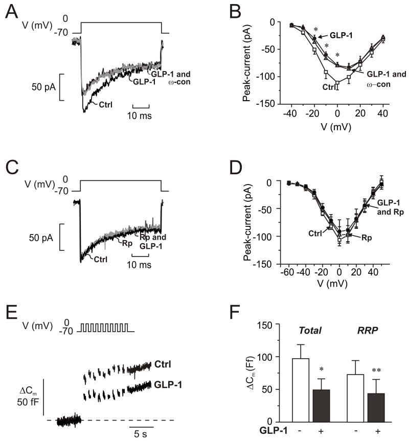
GLP-1 blocks N-type Ca2+-channels and inhibits exocytosis.
(A) Whole-cell Ca2+-currents evoked by membrane depolarization from −70 mV to 0 mV under control conditions (Ctrl; 1 mM glucose), 5 min after addition of GLP-1 (10 nM) and 5 min after addition of 100 nM ω-conotoxin in the continued presence of GLP-1 (GLP-1 and ω-con; grey).
(B) Current (I)-voltage (V) relationship recorded using the perforated patch whole-cell configuration under control conditions (□; n=10), 5 min after addition of 10 nM GLP-1 (●; n=10), and 5 min after addition of ω-conotoxin (100 nM: ω-con) in the continued presence of GLP-1 ( ; n=6). *p<0.05 for effects of GLP-1 vs. control.
(C-D) As in A-B but recorded under control conditions (Ctrl, black), 6 min after the addition of 10 μM 8-Br-Rp-cAMPS (Rp; dark grey) and 4 min after addition of 100 nM GLP-1 in the continued presence of 8-Br-Rp-cAMPS (Rp and GLP-1; light gray) in α-cells
(E) Changes in membrane capacitance (ΔCm) elicited by ten voltage-clamp depolarizations from −70 mV to 0 mV under control conditions (1 mM glucose; □), after the addition of 10 μM 8-Br-Rp-cAMPS  and in the simultaneous presence of 10 nM GLP-1 8-Br-Rp-cAMPS (●).
(F) Histogram of the mean increase in membrane capacitance elicited by the entire train (Total) and increase evoked by the two first depolarizations (RRP). n=6; *p<0.05, **p<0.01.

