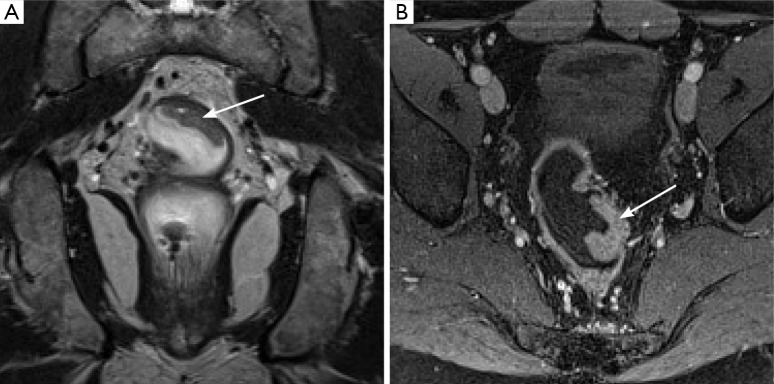
Figure 2.

Example of a T2N0 rectal cancer. Coronal (A) T2 weighted image demonstrates a small polyploid mass (arrow) arising from the wall of the rectum. Importantly, the overlying hypointense line demarcating the muscularis propria remains intact, suggesting this is not a T3 lesion. Axial post-gadolinium image (B) nicely demarcates the mass (arrow), although evaluating extension through the muscularis is not possible on this sequence.
