Fig. 1.
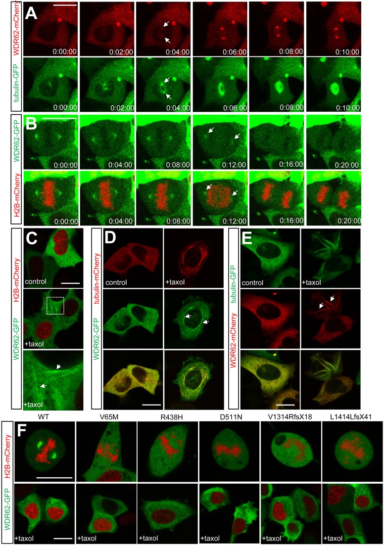
WDR62 associates with microtubules during interphase and mitosis. (A) WDR62–mCherry and tubulin–GFP were coexpressed in AD293 cells and their association with astral microtubules during mitotic entry was revealed by live-cell fluorescence imaging. Arrows indicate spindle association. (B) Decreased spindle pole localization of GFP-tagged WDR62 in cells undergoing metaphase-anaphase transition. Chromosome separation and anaphase transition were determined by H2B–mCherry coexpression. Arrows highlight reduced levels of centrosome-associated WDR62. (C) Taxol treatment (10 µM, 30 min) revealed that WDR62–GFP was partially localized to cytoplasmic filaments (arrows) during interphase. The highlighted area of interest indicated in the middle panel is shown at higher magnification in the lower panel. (D,E) Filamentous WDR62 is colocalized with microtubule bundles (arrows) in taxol-treated non-mitotic AD293 cells. (F) Defective microtubule localization by GFP-tagged MCPH-associated WDR62 missense (V65M, R438H, D511N) and truncated frame-shift (V1314RfsX18, L1414LfsX41) mutants in mitotic or taxol-treated (10 µM, 30 min) interphase cells. Scale bars: 20 µm.

