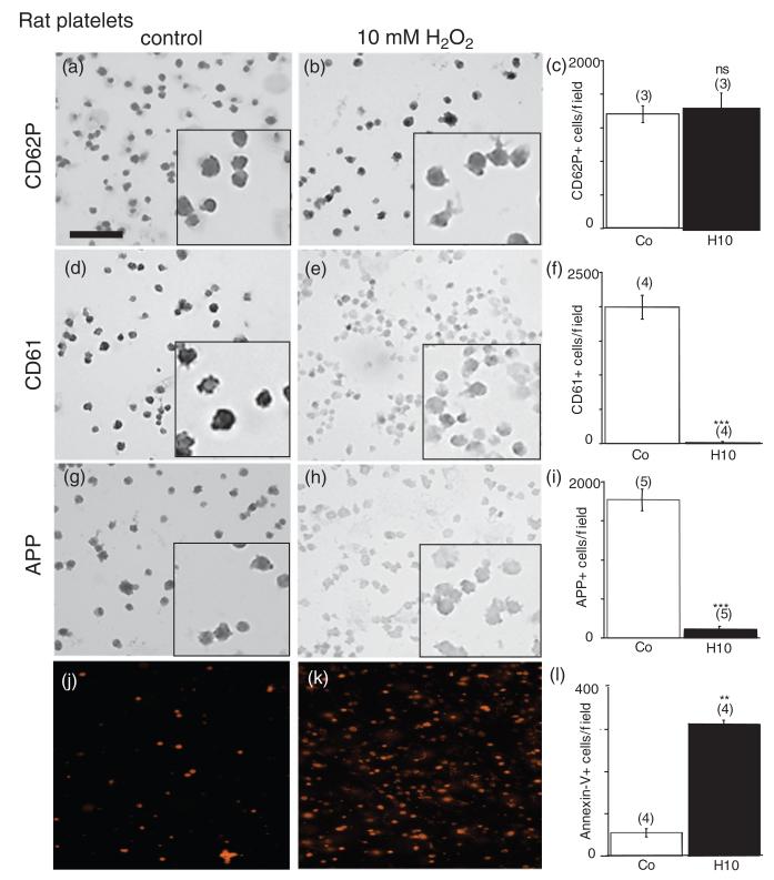
Figure 1.
Immunohistochemical staining of rat platelets. Rat platelets were treated with 10mM H2O2 for 20 minutes (b, e, h, k) or without (a, d, g, j) and spotted onto glasslides. Immunohistochemistry against CD62P (a, b) or CD61 (d, e) or amyloid precursor protein (APP) (g, h) or Annexin-V-staining (j, k) was performed. H2O2 markedly reduced the number of CD61 (e, f) and APP-positive platelets (h, i), but not of CD62P (b, c) and enhanced Annexin-V-positive cells (k, l). Platelets were counted under the microscope at 40 × magnification in a 336 × 448 μm field. Values are given as mean ± SEM. Statistical analysis was performed by Students t-test. Values in parenthesis give the number of experiments. *** p < 0.001;**p < 0.01; ns, not significant; Scale bar in (a) = 24 μm (large image) and 9 μm (small inserts), and 40 μm (j, k).

