Figure 2.
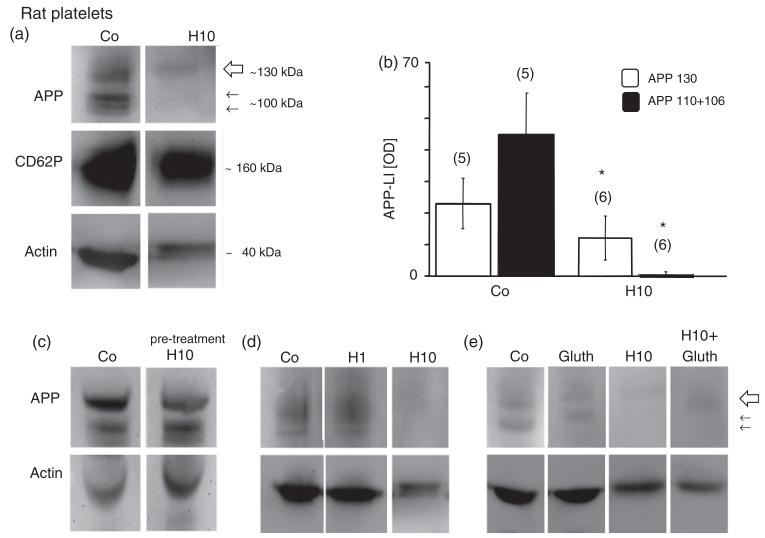
Western blot analysis revealed three bands of amyloid precursor protein (APP) fragments at 130 (large arrow) and 110, 106 (small arrows) kilo Dalton (kD) in rat platelets (a). CD62P was used as a well established marker for control platelets and revealed a size of approximately 160 kDa (a). Actin was used for quantitative correlation and revealed a size of approximately 40 kDa (a) in control rat platelets. Incubation of rat platelets with 10mM H2O2 (H10) dramatically decreased the APP immunoreactivity of all 3 bands after 20 minutes (a), but did not affect CD62P (a). The size of actin was slightly increased after H2O2 treatment (a). H2O2 markedly reduced the APP 130, 110, and 106 kDa fragments, as shown in quantitative analysis (b). Pre-treatment of blots with 10mM H2O2 did not affect immunogenicity of the monoclonal 22C11/APP antibody (c). The effect of H2O2 was dose-dependent (d) and seen at 10mM H2O2 (H10), but not at 1mM H2O2 (H1). Glutathione (Gluth) partly counteracted the H2O2-induced (H10) decline of APP expression (e). Each lane represents an individual subject. Values are given as mean ± SEM optical density (OD). Statistical analysis was performed by students t-test. Values in parenthesis give the number of subjects; *p < 0.05.

