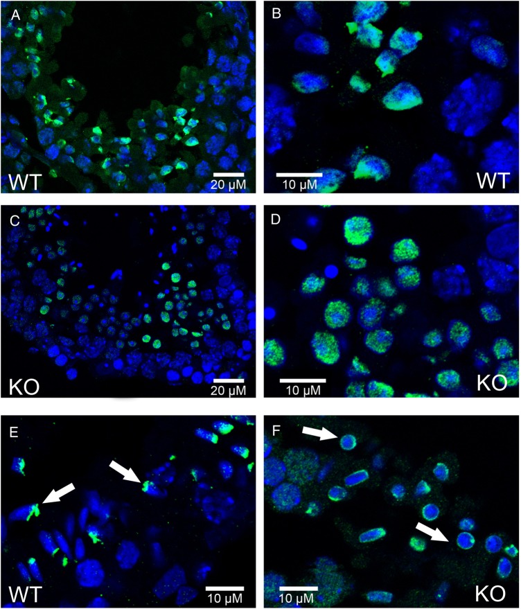
Figure 1.
Histone H4 is acetylated in both WT and Dpy19l2 KO testis sections, but present different pattern of vanishing. (A–F) Testis sections were fixed and stained with Hoechst to mark DNA (blue) and with antibodies targeting acetylated histone H4 (H4ac, green signal). (A and B) Wild type (WT) testis sections showing H4 acetylation on late round spermatids. (C and D) Testis section from Dpy19l2 knock-out (KO) males showing H4 acetylation on late round spermatids. (E) WT testis sections showing the localization of H4ac on elongating spermatids (white arrows). (F) Testis section from Dpy19l2 KO males showing the localization of H4ac on condensed spermatids, which likely corresponded to WT elongated spermatids.

