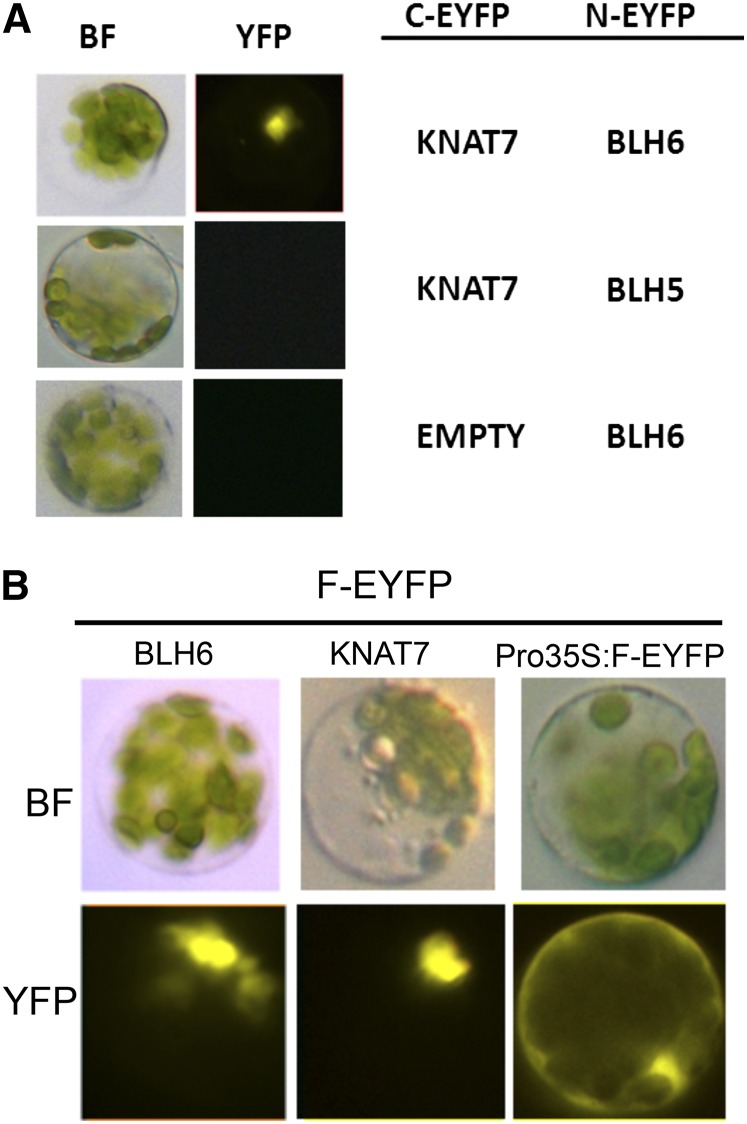
Figure 1.
Bimolecular Fluorescence Assay of KNAT7-BLH6 Interaction and BLH6-EYFP Localization.
(A) Left column: bright-field images of representative protoplasts cotransfected with KNAT7-C-EYFP and BLH6-N-EYFP, KNAT7-C-EYFP and BLH5-N-EYFP, and EMPTY-C-EYFP and BLH6-N-EYFP. Right column: images of the same protoplasts obtained by a Leica DM-6000B fluorescent microscope.
(B) Top row: bright-field images of representative protoplasts transfected with full-length BLH6-EYFP and KNAT7-EYFP fusions, and full-length EYFP alone under the control of the 35S promoter. Bottom row: images of the same protoplasts obtained by the same microscope to image YFP fluorescence.
C-EYFP, C-terminal enhanced YFP protein; N-EYFP, N-terminal enhanced YFP protein; F-EYFP, full-length enhanced YFP protein.

