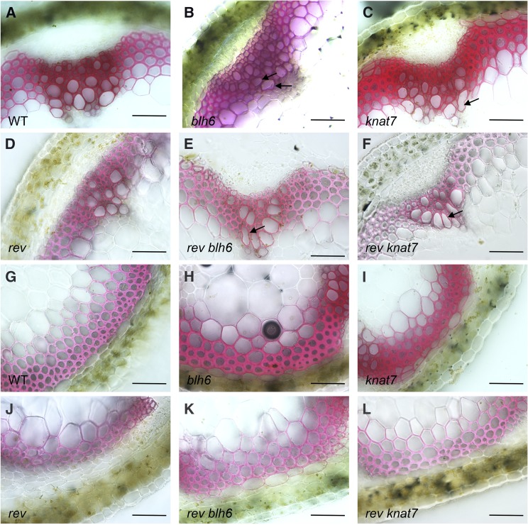
Figure 12.
Inflorescence Stem Secondary Cell Wall Phenotypes in blh6 rev and knat7 rev Mutants.
Stem cross sections were taken 3 cm from the bases of the inflorescence stems of 6-week-old plants, stained with phloroglucinol, and examined for alterations in anatomy and secondary wall thickness. Arrows indicate irx vessel cells. Bars = 50 μm.
(A) and (G) Representative cross sections from wild-type control stems showing vascular bundle (A) and interfascicular fiber (G) regions of the stem.
(B) and (H) Representative cross sections from a blh6 stem showing the vascular bundle region with the irx phenotype (B) and interfascicular fiber region with thicker secondary cell walls relative to the wild type (H).
(C) and (I) Representative cross sections from a knat7 stem showing the vascular bundle region with the irx phenotype (C) and interfascicular fiber region with thicker secondary cell walls relative to the wild type (I).
(D) and (J) Representative cross section from a rev-5 stem showing the vascular bundle region (D) and interfascicular fiber region with thinner secondary cell walls relative to the wild type (J).
(E) and (K) Representative cross section from a rev blh6 stem showing the vascular bundle region with the irx phenotype (E) and interfascicular fiber region (K) with thinner secondary cell walls relative to the wild type and blh6.
(F) and (L) Representative cross section from a rev knat7 stem showing the vascular bundle region with the irx phenotype (F) and interfascicular fiber region (L) with thinner secondary cell walls relative to the wild type and knat7.

