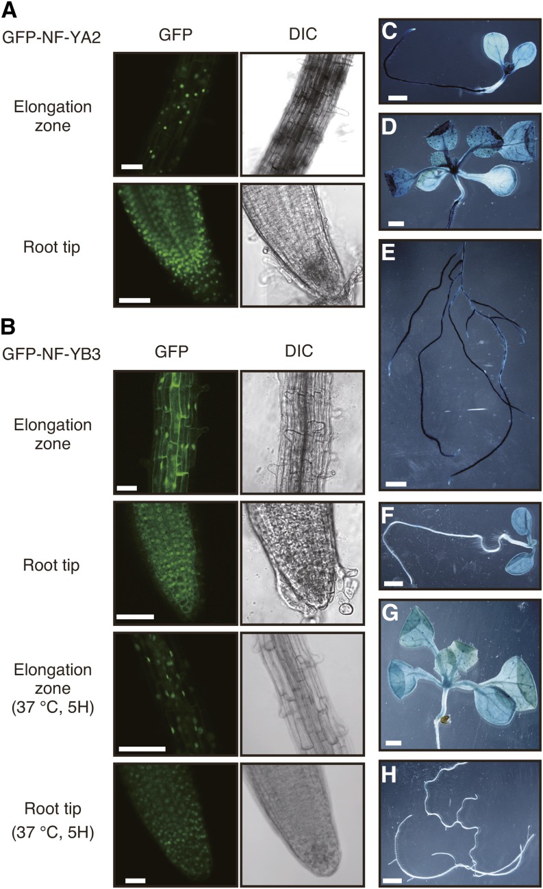
Figure 7.
Subcellular Localization of NF-YA2 and NF-YB3 Subunits and Tissue-Specific Expression Patterns of the NF-YA2 and NF-YB3 Genes.
(A) Nuclear localization of sGFP-NF-YA2. The root tissues of 35S:sGFP-NF-YA2 plants were observed under a microscope. Confocal images of GFP fluorescence and differential interference contrast (DIC) images are shown. Bars = 100 μm.
(B) Nuclear translocation of sGFP-NF-YB3 under heat stress conditions. The root tissues of 35S:sGFP-NF-YA2 plants were observed under a microscope. Confocal images of GFP fluorescence and differential interference contrast (DIC) images are shown. Bars =100 μm.
(C) to (H) GUS staining of NF-YA2pro:GUS ([C] to [E]) or NF-YB3pro:GUS ([F] to [H]) transgenic plants at different growth stages. Whole 7-d-old ([C] and [F]) and aerial ([D] and [G]) and root ([E] and [H]) tissues of 3-week-old seedlings. Bars = 1 mm.

