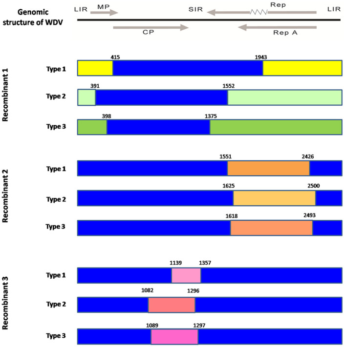
Figure 2. Recombination events detected for the 229 isolates of Wheat dwarf virus and 1 isolate of Oat dwarf virus.
The results showed three recombinants. The genomic structure of WDV was marked by gray color on the top of the figure, in which the mp and cp were on the positive-sense strand, whilst Rep, Rep A and intron were indicated with a zigzag line were on the antisense strand. The three recombinants have three different configurations. Nucleotide sites in the genomic sequence are labeled. Each color represents a different type of recombinant. The blue framework represents the genomic structure of WDV.

