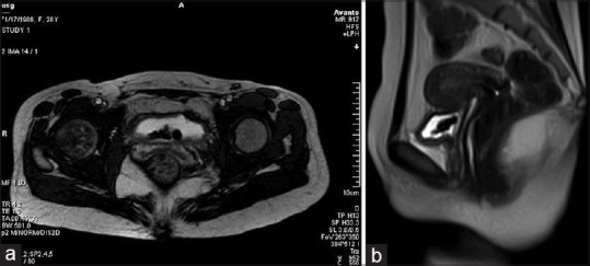
Figure 4.

(a) T2-weighted axial image showing partially distended urinary bladder with hypo intense intrauterine contraceptive devices. (b) T2-weighted sagittal image showing uterus with normal morphology. No fistulous tract seen between uterine bleeding (UB) and uterus. Hypo intense intrauterine contraceptive devices seen in the UB
