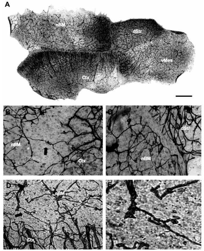
Fig. (3).
Laminin-positive capillaries in a co-slice. Co-slices composed of four brain regions, the basal nucleus of Meynert (nBM), dorsal striatum (dStr), ventral mesencephalon (vMes) and cortex (Ctx) were incubated for 4 weeks with nerve growth factor and glial cell line-derived neurotrophic factor and then stained for laminin (A-E). Higher magnifications of the borders between two brain regions are shown in Fig. (B-E). Note that laminin-positive capillaries cross the border between nBM and Ctx (B, arrow), or are damaged (B). A laminin-negative area between the nBM and dStr is displayed in (C). Co-cultures after thapsigargin treatment displayed an enhanced crossing of laminin-positive capillaries between the nBM and Ctx (D, E, arrow). Fig. (E) showed a higher magnification of vascular growth in Fig. (D). Scale bar in A = 900 μm (A), 300 μm (B-D), 100 μm (E).

