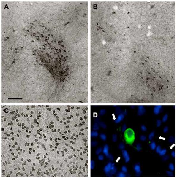
Fig. (4).
Effects of thapsigargin on cell death. Co-cultures were treated overnight without (control, A) or with 3 μM thapsigargin (B-D). After 3 days, slices were immunohistochemically stained against tyrosine hydroxylase (A, B), which stains dopaminergic neurons in the vMes. In situ labelling of nuclear DNA fragmentation exhibited an enhanced TUNEL-positive staining in vMes slices after thapsigargin (C). Co-localization experiments showed malformed DAPI-positive nuclei (blue, arrows), which did not co-localize with TH-positive neurons (green, Alexa-488) after thapsigargin treatment (D). Scale bar in A = 250 μm (A, B), 100 μm (C), 50 μm (D).

