
Figure 2.
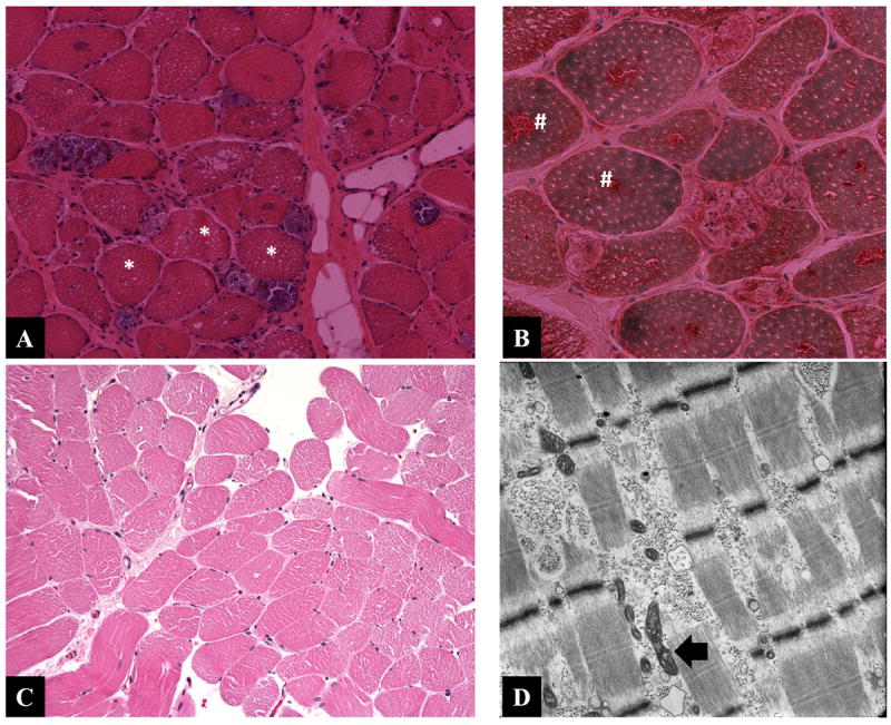
Histological examination of muscle biopsies of patients 9 (A, B) and 4 (C, D). H&E stained muscle biopsy (A) from patient 9 shows extensive vacuolar changes (asterisk) and positive acid phosphatase aggregates (#) in panel B. The H&E stained muscle biopsy (C) from patient 4 is essentially unremarkable while the muscle electron micrograph (D) showed membrane bound glycogen deposits and mildly distorted mitochondrial morphology.