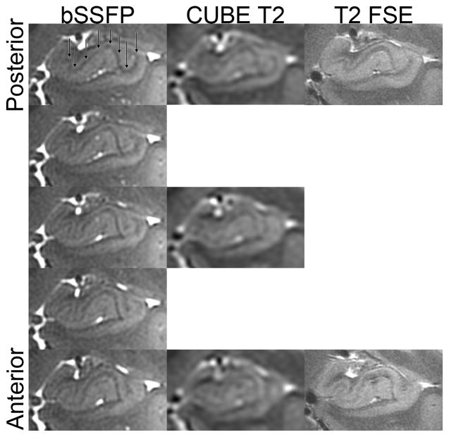
Figure 5.
Comparison of left hippocampal head anatomy on bSSFP, matched posterior-to-anterior sections in a healthy subject. Black arrows point to layer stratum radiatum and lacunosum-moleculare (SRLM [30, 47–49]), the hypointense band centrally within the hippocampus. Images for the CUBE T2 and T2-weighted FSE are displayed in native space without alignment or interpolation.

