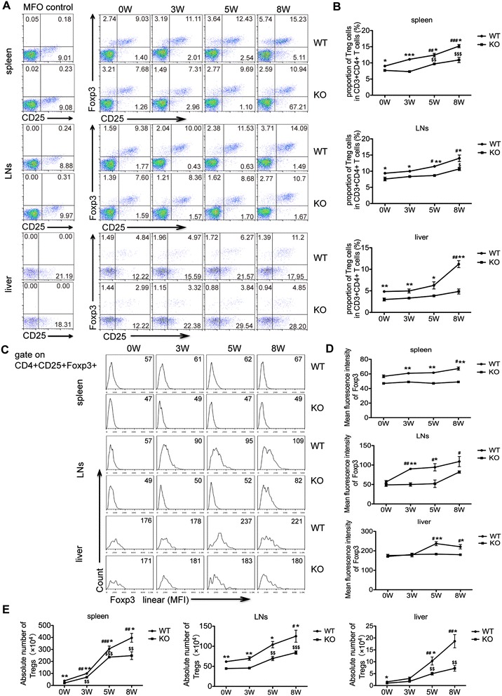
Figure 6.

Treg cells are reduced in S. japonicum- infected AQP4 KO mice . (A) FCM analysis from one representative experiment. At 0, 3, 5, 8 weeks post-infection, four AQP4 WT or KO mice were sacrificed and single cell suspensions of splenocytes, mesenteric lymphocytes or liver cells were prepared for FCM analysis of Treg cells. (B) Proportions of Treg cells in CD3+CD4+ T cells isolated from the spleen, mesenteric lymph nodes, and liver. Representative histograms obtained by FCM analysis (C) of mean fluorescence intensity (MFI) of Foxp3 expression in Treg cells (D). (E) The absolute number of Treg cells in the spleen, lymph nodes or liver from AQP4 WT and KO mice. Data represent means ± SD of 8 mice from two independent experiments. #P < 0.05, ##P < 0.01, ###P < 0.001 vs. AQP4 WT-0 W; $P < 0.05, $$P < 0.01, $$$P < 0.001 vs. AQP4 KO-0 W; *P < 0.05, **P < 0.01, ***P < 0.001 Treg cells from AQP4 KO mice vs. from AQP4 WT mice at 0, 3, 5, 8 weeks post-infection.
