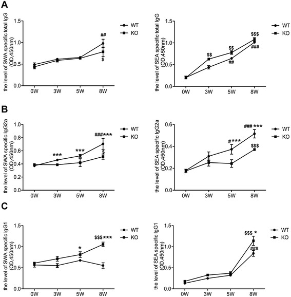
Figure 8.

AQP4 KO mice show higher IgG1 but lower IgG2a levels after S. japonicum infection . At 0, 3, 5, 8 weeks post-infection, four AQP4 WT or KO mice were sacrificed and the serum samples were collected for standard ELISA using the SWA and SEA as the coated antigen. (A) The kinetics of the level of total IgG in the serum from AQP4 WT or KO mouse. SEA and SWA specific IgG2a (B) and IgG1 (C) antibodies in serum from S. japonicum infected AQP4 WT and KO mice were detected by ELISA. Results are expressed as mean ± SD of 8 mice from two independent experiments. #P < 0.05, ##P < 0.01, ###P < 0.001 vs. AQP4 WT-0 W; $P < 0.05, $$P < 0.01, $$$P < 0.001 vs. AQP4 KO-0 W; *P < 0.05, **P < 0.01, ***P < 0.001 total IgG, IgG1 and IgG2a cells from AQP4 KO mice vs. from AQP4 WT mice at 0, 3, 5, 8 weeks post-infection.
