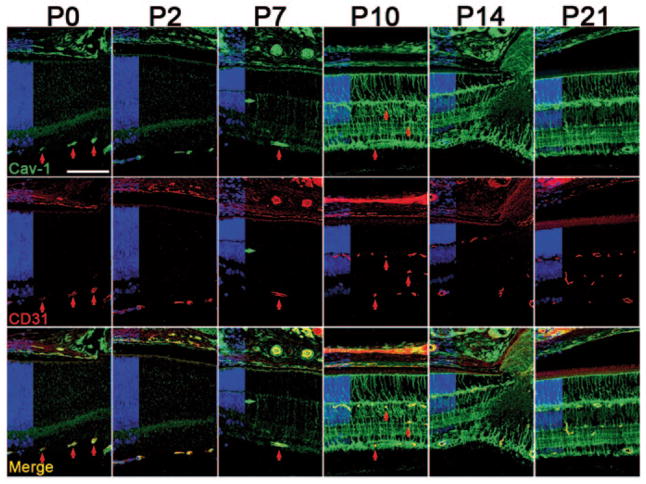
Figure 3.1.
Caveolin-1 (Cav-1) is expressed in developing vessels and increases in the neuroretina from P7–P10. Cav-1 (green) colocalizes with the endothelial marker, cluster of differentiation 31 (CD31, red) in vessels throughout postnatal retinal development. Red vertical arrows highlight several vessels at various developmental stages. The green horizontal arrow at P7 indicates a CD31-negative radial cell. Nuclear layers are indicated in blue on the left of each panel. (Scale bar = 100 μm)

