Figure 7.
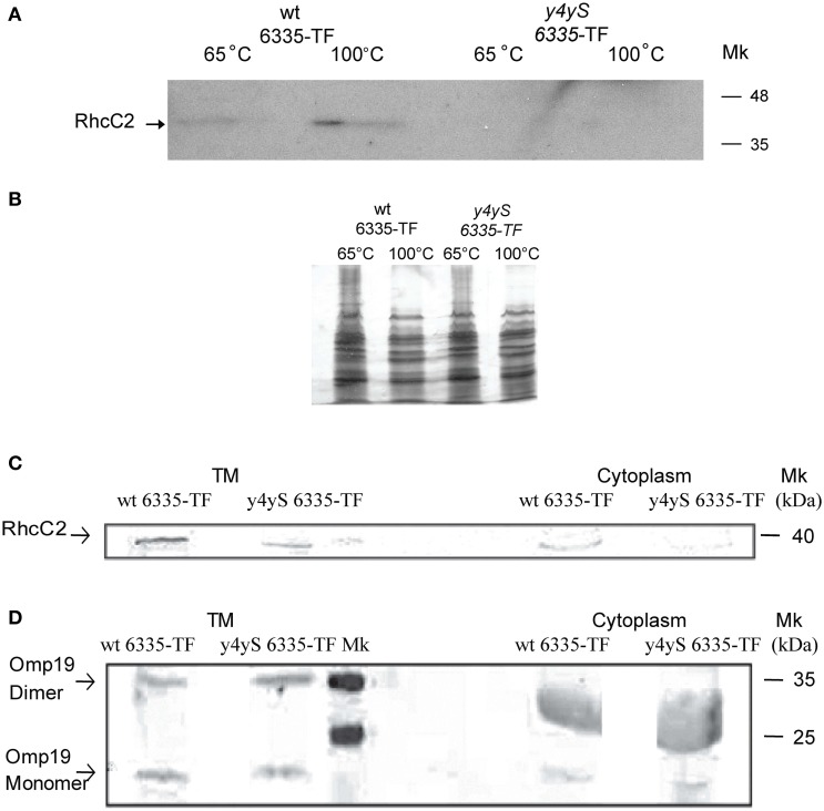
Detection of the 3x FLAG RhcC2 fused protein. (A) OMs of wt 6335-TF and y4yS mutant strain with sequence encoding the 3xFLAG RhcC2 fused protein integrated into the bacterial chromosome (y4yS 6335-TF) heated at 65°C or 100°C were separated by 15% SDS-PAGE and then immuno-bloted and probed with an anti-FLAG antibody, (B) Silver staining of samples described in A separated by 7.5% SDS-PAGE. Total membranes (TM) and cytoplasmic fractions of wt 6335-TF and y4yS 6335-TF strains heated at 100°C, separated by 12.5% SDS-PAGE and then immuno-bloted and probed with anti-FLAG (C) and anti-Omp19 (D) antibodies and revealed with fluorescent antibodies. All bacteria contain plasmid pMP2112 and all bacterial cultures were made in the presence of naringenin. Positions of RhcC2, and of monomer and dimer of Omp19 are indicated. Positions of size markers loaded onto the gels are labeled (in kDa). Anti-Omp19 antibodies nonspecifically probe a great band both in wt and mutant cytoplasmic fractions between markers of 25 and 35 kDa.

