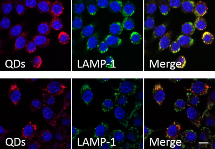
Figure 10.
Colocalization of Qdots and lysosomes. J774 cells were incubated with Qdots (red) for 2 h and fixed afterwards (upper row) or 24 h later (lower row). Cells were immunostained with anti-LAMP1 (detected with Cy2, green). The “Merge” images show that Qdots are localized in lysosomes (yellow). Scale bar: 50 µm.

