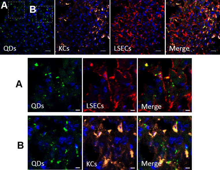
Figure 9.
Confocal microscopy of a cryosection of a rat liver 2h after intravenous injection of polymer-coated Qdots. The nuclei are stained with DAPI. Immunostaining of Kupffer cells (KCs, anti-CD31) and liver sinusoidal endothelial cells (LSECs, with anti-CD68) was performed. Regions outlined by the white boxes are magnified in the lower panels. Nanoparticles can be found located in endothelial cells (A) as well as in Kupffer cells (B), but not in hepatocytes. Scale bar: 20 µm for the upper panel, and 5 µm for the magnified images.

