
Figure 1.
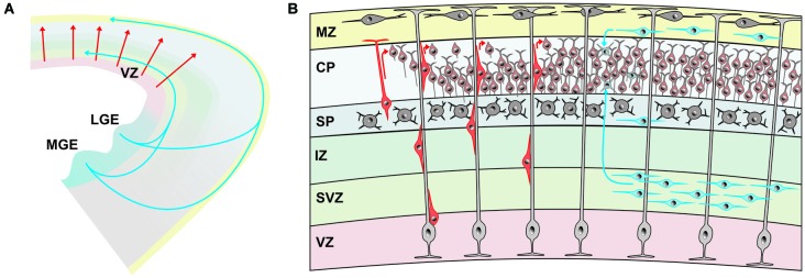
Origin and migratory pathways of glutamatergic and GABAergic neurons. (A) Schematic diagram illustrating migration pathway of the majority of glutamatergic neurons, originating in the ventricular zone (VZ) of the pallium and radially migrating into the developing cerebral cortex (red arrows). The majority of GABAergic neurons are generated in the medial (MGE) and lateral gangionic eminence (LGE) and reach their final position by tangential migration via deep pathways and superficial cortical layers. (B) Glutamatergic neurons (marked in different shades of red) are generated in the VZ and migrate radially either by somal translocation or, at later phases, by locomotion along radial glial cells (light gray). Upon reaching the marginal zone (MZ) they detach and align on top of previously generated neurons of the cortical plate (CP), generating the “inside first—outside last” pattern of the cerebral cortex. The majority of GABAergic neurons (marked in different shades of blue) reach the cortex via tangential migration in the deep pathway within the subventricular zone (SVZ) or the superficial pathway in the MZ. Some GABAergic interneurons travel also within the subplate (SP).