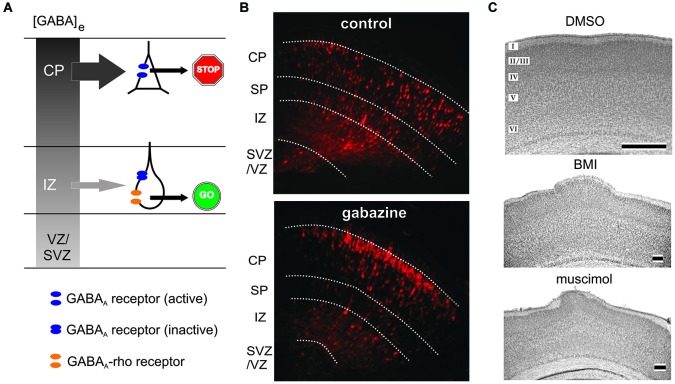
Figure 4.
Role of ionotropic GABA receptors on radial migration. (A) Model of GABAA and GABAA-rho receptor dependent radial migration in the neonatal cerebral cortex, which shows outside directed GABA gradient (gray colored gradient). In the IZ migrating neurons express functional GABAA receptors (blue discs) and GABAA-rho receptors (orange discs), whereas in the CP migrating neurons express only functional GABAA receptors. Due to the outside directed GABA gradient the low-affinity GABAA receptors are only activated in the CP, while the lower GABA concentration in the IZ is sufficient to activate the high affinity GABAA-rho receptors. Activation of GABAA-rho receptors is necessary to support migration in the IZ (GO sign), while activation of GABAA receptors contributes to termination of migration (STOP sign). (B) Blockade of GABAA receptors with gabazine facilitates radial migration. Figures illustrate red fluorescent protein (RFP)-positive cells in control (left) and gabazine-treated (right) GAD67GFP/GFP fetuses at E17.5, which were injected with gabazine at E14.5 immediately after the electroporation of the RFP vectors. (C) Digital photographs of Nissl-stained coronal sections from a P7 rat that received at P0 on the cortical surface an Elvax implant containing DMSO (top, control), the GABAA antagonist bicuculline methiodide (middle, BMI) or the GABAA agonist muscimol (bottom). Note upper layer heterotopia due to increased radial migration in BMI- and muscimol-treated animals. Scale bar in B, C middle and C bottom corresponds to 200 µm, in C top to 500 µm. Modified with permission from Denter et al. (2010) (A), Furukawa et al. (2014) (B), and Heck et al. (2007) (C).

