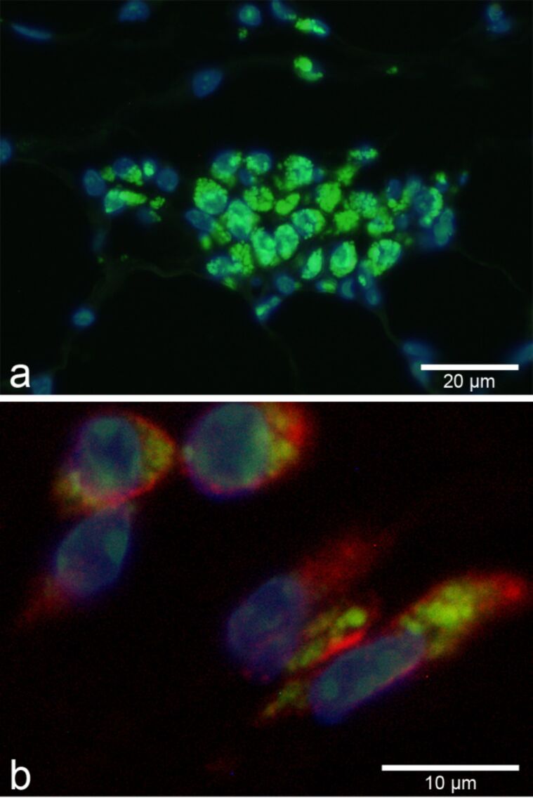
Figure 3.
Aggregates of FITC-labeled SiO2-NP (green, 55 ± 6 nm in diameter) were visualized by fluorescence microscopy in macrophages following subcutaneous injection (a). Nuclei were counterstained with DAPI (blue). Subsequent immunofluorescent labeling (red) for the macrophage marker F4/80 identified macrophages as uptaking cells (b).

