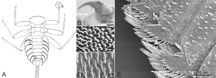
Figure 6.
(A–D) Attachment devices of E. assimilis larvae: (A) ventral view, (B) claw of the foreleg, (C) setae of the pads on the ventral side of the gill lamellae, (D) areas with spiky acanthae on the lateral parts of the abdominal sternits. Reproduced with permission from [26]; (E) Structure on the distal edge of the ventral side of the beetle larva Elmis sp. Reproduced from [18].

