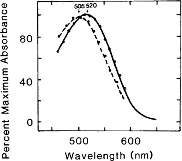
Fig. 3.

Difference spectra of porphyropsin and isoporphyropsin regenerated from the incubation of 11-cis and 9-cis isomers of 3,4-didehydroretinal with bleached rod outer segments of goldfish. Purified rod outer segments were completely bleached (room light, 10 min) before isomers of 3,4-didehydroretinal (13-cis, 11-cis, 9-cis, and all-frans) were added to them (for details, see Methods). Only the 11-cis and the 9-cis isomers resulted in the regeneration of visual pigments. The former regenerated the native goldfish porphyropsin (solid line, absorbance maximum: 520 nm) whereas the latter regenerated the isoporphyropsin (broken line, absorbance maximum: 505nm). The open and solid circles are nomogram values for these porphyropsins (Ebrey and Honig, ’77).
