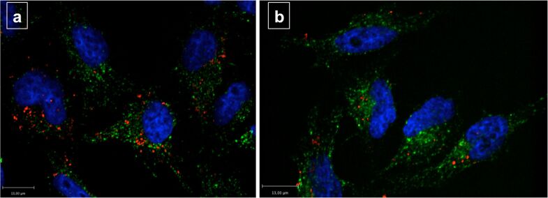
Figure 7.
Fluorescence image of Hela cells which were incubated with SCIONs (iron concentration 5 µg/mL, incubation time 4 h), blue = DAPI (nuclei), green = Transferrin Alexa Fluor® 488 conjugate (cytosol), red = Alexa Fluor® 555 (SCIONs); (a) Control cells without siRNA treatment; (b) Cells with knockdown of Caveolin-1.

