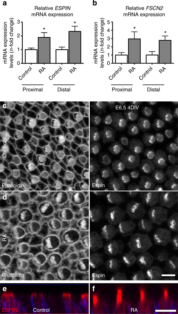
Figure 8. RA treatments upregulate expression for actin crosslinking proteins expressed within stereocilia.
(a) Relative mRNA expression levels for Espin in proximal and distal regions of E6.5 BPs cultured 3 days in control medium or medium supplemented with RA. Expression levels were normalized relative to the average mRNA expression in vehicle controls. Espin expression was 1.9-fold greater in the proximal region with RA treatments (P= 0.0147; Student’s t-test; n= 5) and 2.3-fold greater in the distal region (P = 0.0079, Student’s t-test; n = 7). (b) Relative mRNA expression levels for Fscn2 in proximal and distal regions of E6.5 BPs cultured for 3 days in control medium or medium supplemented with RA. Expression levels were normalized relative to the average mRNA expression in vehicle controls. Fscn2 expression was threefold greater in the proximal region with RA treatments (P = 0.0295, Student’s t-test; n = 3) and 2.8-fold greater in the distal region (P = 0.0305; Student’s t-test; n = 4). (c) Proximal HCs that developed in E6.5 BPs from the left ear that were cultured for 4 days in vehicle control medium, fixed and immunolabelled with Espin. Left panel shows the grey scale from Alexa Fluor-488-phalloidin labelling and the right panel shows the grey scale image from Espin labelling. HCs formed hair bundles with circular outlines. (d) Proximal HCs that developed in E6.5 BPs from the right ear that were cultured for 4 days in medium supplemented with RA. Hair bundles in these HCs formed semicircular outlines with more pronounced Espin immunofluorescence. (e,f) Images showing z axis of Espin (red) and HCS-1 (blue) merged immunofluorescence in proximal HCs of E6.5 BPs cultured 4 for days with vehicle control (e), or with RA (f). The Espin-positive hair bundles were noticeably longer in the HCs that were exposed to exogenous RA. Graphs show mean mRNA expression levels (n-fold change) ±s.e.m. Scale bars, 5µm.

