Fig. 5.
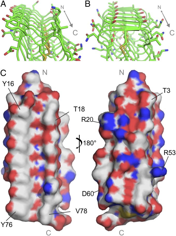
The midge AFP has ice-binding residues not observed in other AFPs. (A) Predicted ice-binding site (IBS) of midge AFP. (B) IBS of TmAFP. The C-terminal ends are to the front in A and B. (C) Surface representation of the midge AFP model showing the putative flat ice-binding surface (Left) and the opposing charged, uneven surface (Right). The N and C termini are indicated in gray.

