Fig. 2.
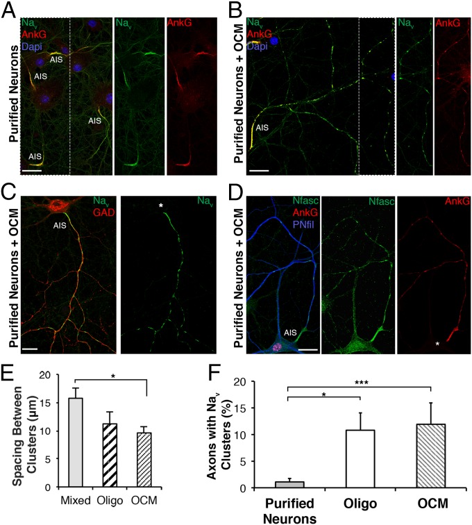
A secreted oligodendroglial factor promotes nodal protein clustering. Immunostaining of purified hippocampal neurons cultured in the (A) absence or (B–D) presence of OCM. (A–D) The AIS is detected in all conditions, but (B–D) clusters of Nav (green), AnkG (red), and Nfasc (green) are only detected in the presence of OCM. (C) Prenodes are formed on GAD67+ neurons treated with OCM. (D) Neuronal cell body and neurites stained with an Ab targeting phosphorylated neurofilaments (PNfil; blue). (Scale bars: 25 µm.) (E) Distance between prenodes measured on axons in mixed culture or purified neurons cocultured with oligodendrocytes (oligo) or OCM. Mean spacing between clusters (micrometers) ± SEM of four independent experiments is shown. *P = 0.028 (Mann–Whitney test). (F) Percentage of axons (AIS+) having at least two Nav clusters at 21 DIV in purified neuron cultures, purified neurons cocultured with oligodendrocytes (oligo), or OCM. The means ± SDs of 4 (for oligo) and 10 (for OCM) independent experiments are shown. For each experiment, at least 100 neurons were analyzed. *P = 0.0187; ***P = 0.0003 (Mann–Whitney test).

