Fig. 3.
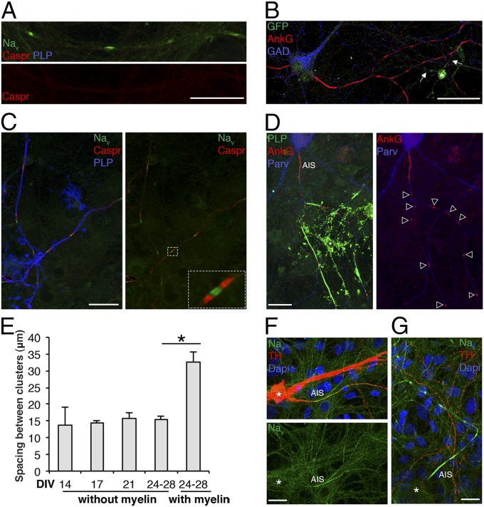
Prenodes are formed on myelin-competent GABAergic neurons. (A and B) Immunostaining of mixed hippocampal culture at 17 DIV. (A) Nav clusters (green) observed in the absence of Caspr aggregation (red) and the absence of myelin (PLP−; blue). (B) Hippocampal culture from CNP-EGFP embryos shows an oligodendrocyte (asterisk) stained with an anti-GFP Ab with processes contacting the axon (arrows). Numerous AnkG clusters (red) are observed at a distance from these processes on a GAD67+ neuron (blue). (C and D) Immunostaining of mixed hippocampal culture at 24 DIV. (C) Myelinated axons (PLP+; blue) with nodes (Nav; green) and paranodes (Caspr; red). A higher magnification of a node with flanking paranodes is shown. (D) Myelinated segments (PLP+; green) with nodes (AnkG+; red) on GABAergic axons [Parvalbumin+ (Parv); blue]. (Scale bars: 25 µm.) (E) Measure of the distance between prenodes on unmyelinated axons and between nodes of Ranvier on myelinated axons at different time points. Values are the mean spaces between clusters (micrometers) ± SEMs of four independent experiments. *P = 0.028 (Mann–Whitney test). (F and G) Immunostaining of mesencephalic cultures at 21 DIV. (F) Dopaminergic neurons tyrosine hydroxylase (TH+; red) do not form Nav clusters downstream of the AIS (Nav+; green), whereas (G) Nav clusters are formed on a nondopaminergic (TH−) axon. *Neuronal cell body. (Scale bars: 25 µm.)

