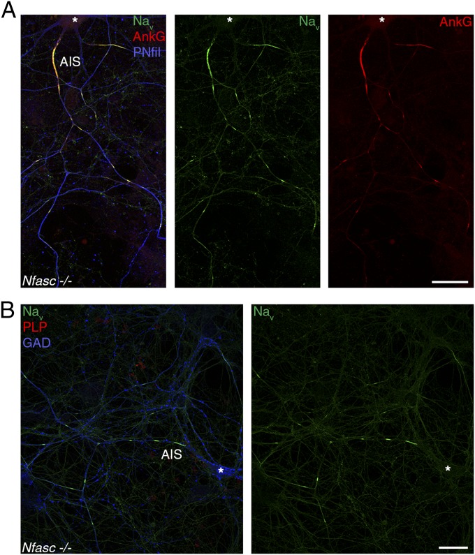
Fig. 5.
Prenodes are formed in the absence of Nfasc expression. Immunostaining of hippocampal neuron cultures of Nfasc−/− mice at 20 DIV showing clusters of Nav (green) and AnkG (red) along an axon with phosphorylated neurofilaments (PNfil; blue) in A and in the absence of myelin (PLP−; red) on a GAD67+ neuron (blue) in B. *Neuronal cell body. (Scale bars: 25 µm.)

