Fig. 6.
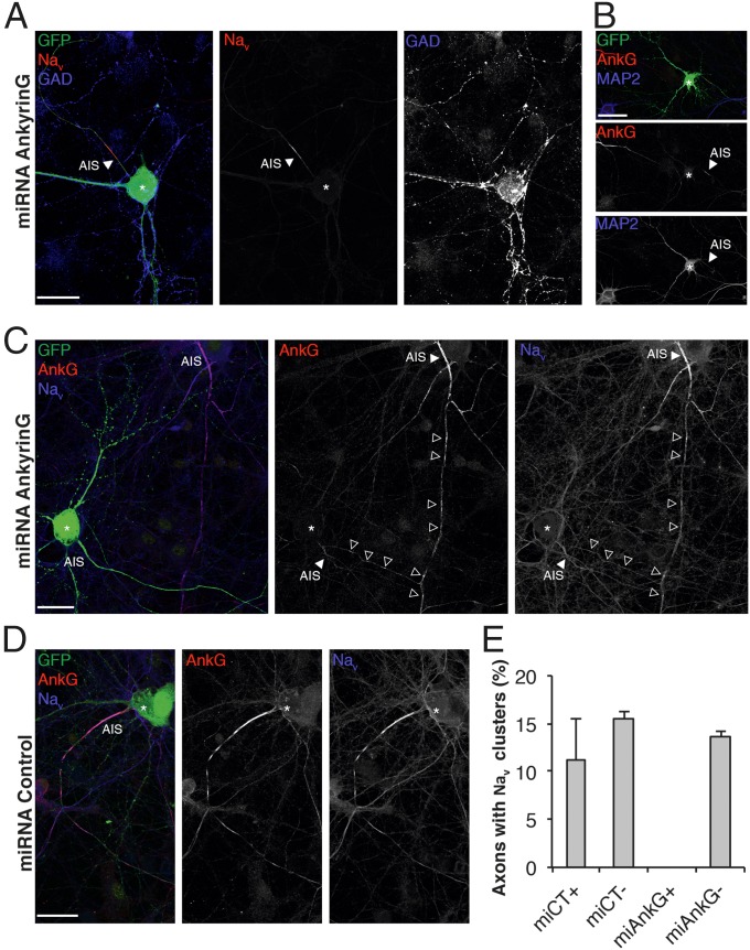
Prenode assembly requires AnkG expression. (A–C) Immunostaining of hippocampal cell cultures transfected at 6 DIV with AnkG miRNA and fixed at 18 DIV. Transfected neurons are GFP+ (green). (A) Representative image of a transfected GAD67+ neuron (blue); Nav expression (red) is weak at the AIS (arrowheads), and no prenodes are observed. (B) Transfected AnkG miRNA GFP+ neuron with somatodendritic expression of MAP2 (blue) and a weak AnkG (red) staining of the AIS (arrowheads). (C) Transfected AnkG miRNA GFP+ neuron with weak expression of AnkG (red) and Nav (blue) at the AIS and no prenodes, whereas prenodes are observed in the neighboring nontransfected neuron (arrowheads). *Neuronal cell body. (D) Image of a neuron transfected with control miRNA showing AnkG (red) and Nav (blue) expression at the AIS and prenodes. (E) Quantification of axons forming Nav clusters in hippocampal cell cultures transfected (+) or not (−) with control miRNA (miCT) or AnkG miRNA (miAnkG). The means ± SDs of three independent experiments are shown. For each experiment, at least 50 neurons were analyzed. (Scale bars: 25 µm.)

