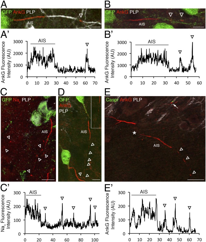
Fig. 8.
Prenodes can be detected in vivo during postnatal development in both mouse and rat. (A and B) Immunostaining of sagittal sections of P13 VGAT-venus rat hippocampus shows the presence of AnkG+ nodes of Ranvier on myelinated fibers (PLP+) in A as well as AnkG axonal clusters in absence of myelin (PLP−) in B on some GABAergic neurons stained with an anti-GFP Ab. (Scale bars: 10 μm.) Intensity profiles corresponding to AnkG expression in (A′) myelinated and (B′) nonmyelinated fibers. Arrowheads indicate AnkG axonal clusters. (C and D) Immunostaining of sagittal sections of P12 GAD-GFP mouse hippocampus shows the presence of (C) Nav and (D) AnkG clusters (arrowheads) in the absence of myelin (PLP−). In D, the boxed area is shown as a zoomed-in view. (Scale bars: 10 μm.) (E) Immunostaining of P12 GAD-GFP hippocampus shows that Caspr is not clustered around prenodal AnkG before myelination (arrowheads), whereas it is accumulated at hemiparanodes flanking nodal AnkG in myelinated fibers (arrow). *Neuronal cell body. (Scale bar: 20 μm.) (C′ and E′) Intensity profiles corresponding to (C) Nav staining and (D) AnkG show isolated peaks corresponding to prenodes (arrowheads).

