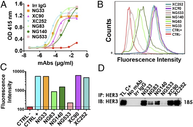
Fig. 1.
A set of mAbs specifically targeting HER3. (A) The 96-well plates were coated by using a solution containing IgB3 (1.5 µg/mL) and thereafter incubated for 1 h with the indicated mAbs. After washing, a second 1-h-long incubation with HRP-labeled anti-mouse IgG was performed and followed by incubation for 20 min with 2,2′-azino-bis(3-ethylbenzothiazoline-6-sulfonic acid). Light absorbance (415 nm) signals are presented. (B and C) NIH/3T3-R2R3 cells were incubated for 60 min at 4 °C with the indicated mAbs (each at 10 µg/mL). After two washes, the cells were similarly incubated with a secondary anti-mouse IgG Ab coupled to Alexa Fluor 488. Fluorescence intensity (F.I.) signals were measured by using the LSRII flow cytometer. Positive and negative controls were used for reference. (D) Protein G beads were incubated first with the indicated mAbs (5 µg; for 2 h at 4 °C), and after two washes, the beads were incubated with cleared N87 cell lysate. After four washes, the immunoprecipitated (IP) proteins were separated by using electrophoresis and immunoblotting (IB) with an Ab to HER3/ERBB3.

