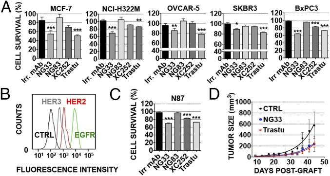
Fig. 4.
Anti-HER3 mAbs decrease NRG-induced tumor cell survival, both in vitro and in animals, as effectively as trastuzumab. (A and C) Proliferation assays using MTT were performed on five different cell lines, as indicated. Cells (5,000 per well) were plated the day before and treated for 72 h with the various agents (each at 10 µg/mL) in medium supplemented with NRG (10 ng/mL). Trastuzumab (Trastu) indicates a huminazed mAb to HER2/ERBB2. (B) N87 cells were incubated for 1 h at 4 °C with mAbs (each at 10 µg/mL) directed to EGFR (565), HER2 (L26), or HER3 (XC252). After two washes, the cells were incubated for 1 h at 4 °C (in the dark) with a secondary anti-mouse Ab coupled to Alexa Fluor 488. Fluorescence intensity (F.I.) was measured by using the LSRII flow cytometer. N87 cell survival was determined as in A. (D) CD1-nude mice were grafted s.c. with 5 × 10⁶ N87 cells. Once tumors became palpable (after ∼13 d), the mice were randomized into group of six animals and treated twice a week for 5 wk. The control group (CTRL) was injected intraperitoneally (IP) with saline (200 µL). The other groups were treated with mAbs at the final concentration of 0.2 mg/0.2 mL of saline per mouse. The mice were weighed once a week, and the tumors were measured twice a week. The average tumor size measured in six mice (± SEM) is shown. ***P < 0.001; **P < 0.01 (ANOVA and post hoc tests).

