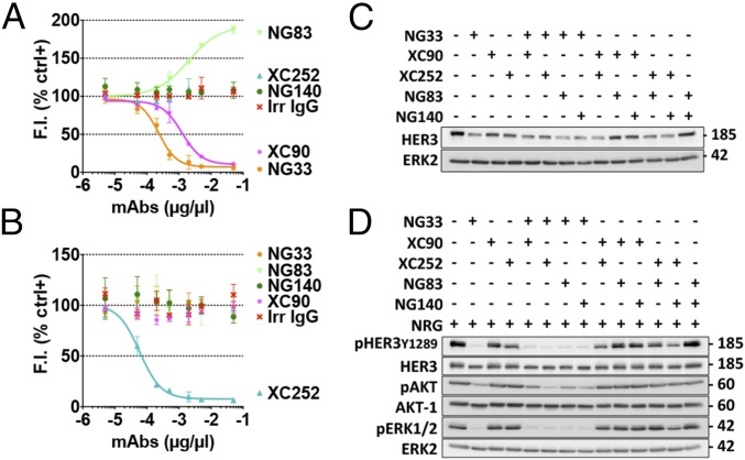
Fig. 5.
Pairwise applications of Abs directed at distinct epitopes of HER3. (A and B) The Abs NG33 and XC252 were labeled with the fluorescent dye Lumi4 Tb Cryptate (K2). The 96-well plates were coated with IgB3 (1.5 µg/mL) and incubated for 1 h with various concentrations of mAbs. The labeled mAb, NG33-K2 (A) or XC252-K2 (B), was then added at 1 nM final concentration. Fluorescence intensity (at 610 nm) was measured after an hour-long incubation. (C) The indicated combinations of anti-HER3 mAbs were studied for their ability to trigger HER3 degradation using N87 cells. Cells were treated for 2 h at 37 °C with mAbs (10 µg/mL). Protein samples were subjected to immunoblotting by using the indicated Abs. (D) The combination’s capacity to modulate NRG-induced phosphorylation of HER3, AKT, and ERK was evaluated by using N87 cells. After 20 min of treatment at 37 °C with the indicated mAbs (10 µg/mL), NRG (20 ng/mL) was added to the cells and incubated for 10 min. Thereafter, the cells were lysed, and equal quantities of lysate proteins were electrophoresed before immunoblotting, as indicated.

