Fig. 2.
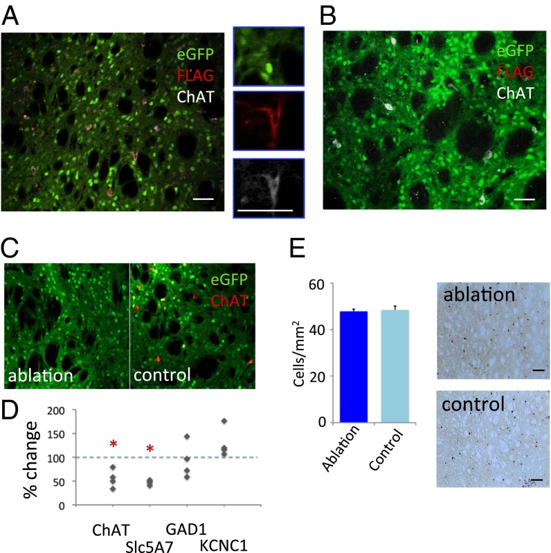
Targeted interneuronal ablation in the ChAT-cre mouse striatum. (A) Triple immunostaining of striatal neurons after A06 virus infusion shows eGFP (green), FLAG (red), and ChAT (white); FLAG immunoreactivity, which corresponds to DTR expression, colocalizes specifically with ChAT, confirming the specificity of DTR expression using this system (compare with D). (B) Triple immunostaining of striatal neurons after C06 virus infusion, showing eGFP (green), FLAG (red; no staining is apparent, although conditions were identical to A), and ChAT (white). (C) Reduced ChAT-expressing interneurons after A06 infection and DT injection in the dorsal striatum, compared with control C06 virus (Fig. 3 B and E). (D) The specificity of cholinergic cell ablation was evaluated using quantitative PCR (qPCR) analysis of RNA isolated from A06-infected and contralateral C06-infected striatum. Expression of ChAT interneuron-related genes was reduced approximately twofold on the ablation side (n = 4 animals. Paired t test: ChAT, P = 0.03 (one tailed); Slc5A7, P = 0.01 (one tailed). GABA-related genes were not significantly reduced: GAD1, P = 0.58 (two tailed); KCNC1, P = 0.054 (two tailed). (E) Immunostaining for parvalbumin-positive interneurons revealed no qualitative or quantitative difference between experimental conditions, further confirming the specificity of our manipulation to the cholinergic interneurons. *P < 0.05. (Scale bar, 20 µm for A–C and 100 µm for E.)

