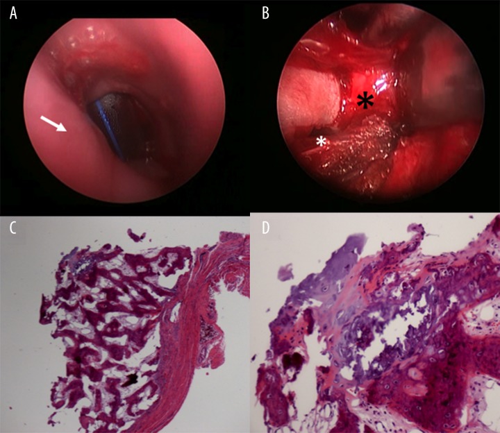
Figure 3.
Direct Laryngoscopy (A). Smooth subglottic mucosa, slight medial displacement of left subglottic wall, immediately lateral to the drained abscess pocket (arrow). In the distance, the tracheotomy tube is seen. Neck Exploration (B). Exposed left anterolateral aspect of cricoid lamina (black asterisk) showing an obvious defect within the wall (white asterisk), in communication with abscess pocket. Subglottic mass lining biopsy (C). Fibroconnective tissue with an attached nodular portion of bone and chondroid tissue (10×). (D) Transition of benign chondroid/cartilaginous and osseous/bony tissue favoring a benign chondro-osseous metaplastic process (40×).

