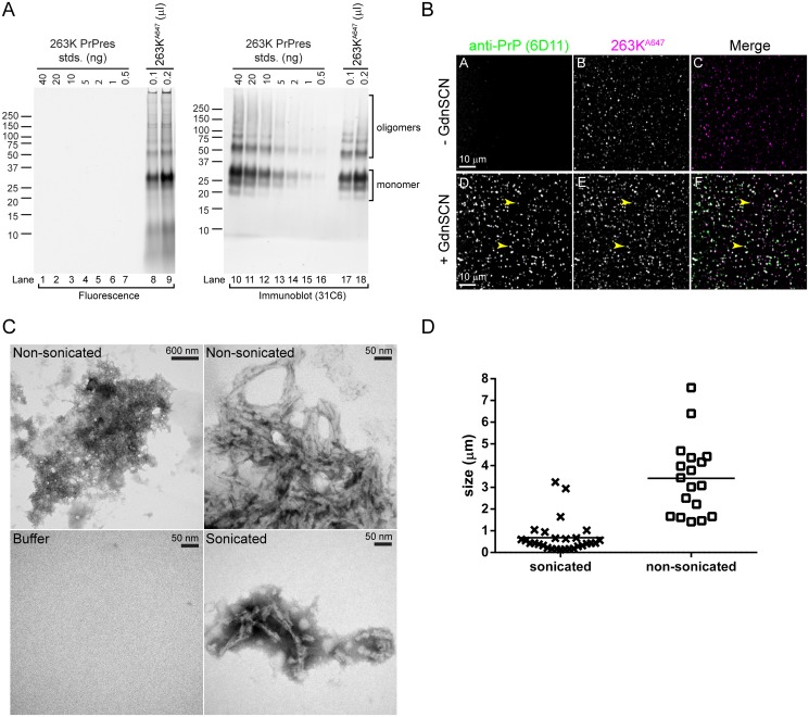
Fig 1. Characterization of 263K PrPres preparations covalently labeled with Alexa Fluor 647 (263KA647).
(A) Fluorescent bands in 263KA647 are PrP. 263KA647 was resolved by SDS-PAGE. The gel was scanned to detect the fluorescent bands (Fluorescence) before immunoblotting with anti-PrP 31C6 (Immunoblot). Known quantities of unlabeled 263K PrPres were loaded as standards (lanes 1–7 and 10–16) for immunoblot quantitation of 263KA647. Fluorescent bands corresponding to 31C6-immunoreactive species constituted >85–90% of the total fluorescence. (B) Fluorescent particles in 263KA647 preparations all contain PrPres. Sonicated 263KA647 was spotted onto coverslips and immunostained with anti-PrP 6D11 with or without pretreatment with 3 M GdnSCN. All fluorescent 263KA647 particles (magenta) exhibited GdnSCN-dependent immunolabeling (green) indicative of PrPres. Merge, white indicates co-localization with equal signal from each channel. Arrowheads indicate examples 263KA647 particles with co-localized immunolabeling. (C) Electron micrographs of 263KA647 aggregates. Samples of 263KA647 before (Non-sonicated) and after (Sonicated) sonication were examined by TEM. Images of a non-sonicated aggregate acquired at low and high magnification are shown to allow visualization of the entire aggregate (upper left) and its composition (upper right). Buffer, buffer only control. (D) Analysis of 263KA647 aggregates in (C). The longest dimension of randomly selected aggregates in TEM images was measured and plotted. The mean size ± standard deviation for sonicated and non-sonicated 263KA647 aggregates was 0.683 μm ± 0.748 μm (n = 28) and 3.415 μm ± 1.676 μm (n = 18), respectively. These differences were statistically significant (P < 0.0001).

