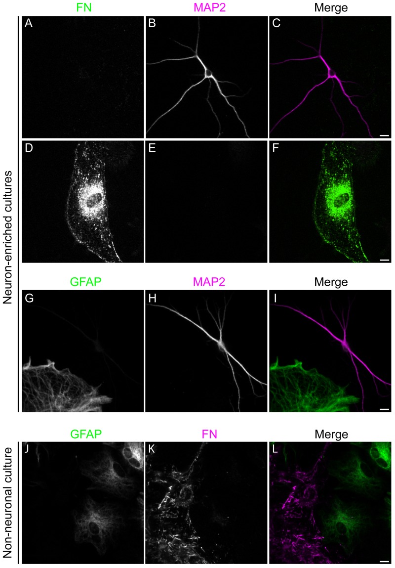
Fig 2. Identification of cell types isolated from adult Syrian hamster brain.
Primary cells isolated from the brain of an adult Syrian hamster were cultured in vitro in NABG (Neuron-enriched) or DMEM+ (Non-neuronal), fixed and then stained for the presence of marker proteins for specific cell types. Neuron-enriched cultures were co-stained for the presence of the neuronal marker protein MAP2 (magenta) and the fibroblast marker fibronectin (FN, green) (shown in A-F) or MAP2 and the astrocyte marker protein GFAP (shown in G-I). Non-neuronal cultures were double stained for the presence of GFAP (green) and FN (magenta) (shown in J-L). Bar = 10 μm.

