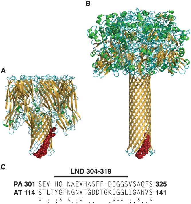
Fig 1. Alpha toxin and protective antigen share structural and functional homology but only limited sequence identity.
Comparison of protein structural models of the heptameric AT (PDB7AHL) (A) and PA (PDB1V36) (B). Sequences in red represent aligned sequence shown in (C). Amino acid alignment of PA and AT in the region of the LND of PA demonstrates 37% sequence identity. Asterisks denote positions of amino acid identity, while periods and colons denote semi-conservative and conservative substitutions, respectively.

